This article is for general MRI education only. It does not replace formal MRI safety training, facility SOPs, accreditation requirements, manufacturer Instructions for Use, or clinical judgment.
An MRI workflow is engineered for consistency: timed appointments, defined protocols, sequence optimization, standardized coils, safety procedures, and quality checks. Yet not every patient fits the equipment, positioning approach, or time allotted for the exam.
For many patients, positioning is straightforward. For others, technologists may need to use manual lifting, improvised support, and repeated adjustments to achieve and maintain position. These arrangements can compress, shift, or contribute to motion artifacts—creating effects that extend far beyond the table.
Too often, those effects are treated as normal operational variation. Technologists see the connection firsthand: when positioning depends on manual lifting, improvised support, or repeated adjustment, the impact can carry through the MRI workflow.
People outside Zone IV may see delayed starts, repeat imaging, overtime, or staff strain without seeing the positioning challenges that contribute to them. As a result, these needs can remain invisible in operational planning.
Connecting cause and effect gives leaders a clearer basis for decisions about appointment length, scheduling, staffing, and equipment. It creates an opportunity to reduce workflow friction, support technologist well-being, and improve patient experience.
Zone IV Needs Reliable Post-Transfer Lifting and In-Bore Support
A collaborative walk-through at each MRI scanner can make these patterns visible to people outside the technologist role, including imaging directors and patient handling, mobility, safety, and risk teams. Invite technologists to share what they encounter, observe how positioning is managed, and identify where variation occurs. Technologists know what they need to perform their work safely and effectively; their experience should help shape the solution.
What Do MRI Clinics Need—and Why Is Today Different?
Technologists are not the only people affected by the changes shaping MRI today. Clinics are being asked to deliver more with less margin for variation. Demand is rising as patient populations grow older and more medically complex. At the same time, staffing shortages, budget constraints, growing exam volumes, and technologies such as AI-enabled workflow tools and remote scanning are reshaping imaging operations.
Accessibility is also part of the operational conversation. The HHS Section 504 Final Rule strengthens requirements for accessible medical diagnostic equipment and for HHS-funded programs to provide nondiscriminatory access to care for people with disabilities. Imaging facilities should evaluate how patients access diagnostic equipment, document their process, and identify barriers to safe, reliable positioning. See HHS Section 504 guidance.
MRI has responded to many pressures by standardizing protocols, safety practices, scheduling, and quality processes. Patient positioning, however, can still depend on manual techniques and improvised solutions when a patient does not fit the expected setup.
That raises an important question: What is non-standardized positioning, and why has it remained a variable while so much else in MRI has become standardized?
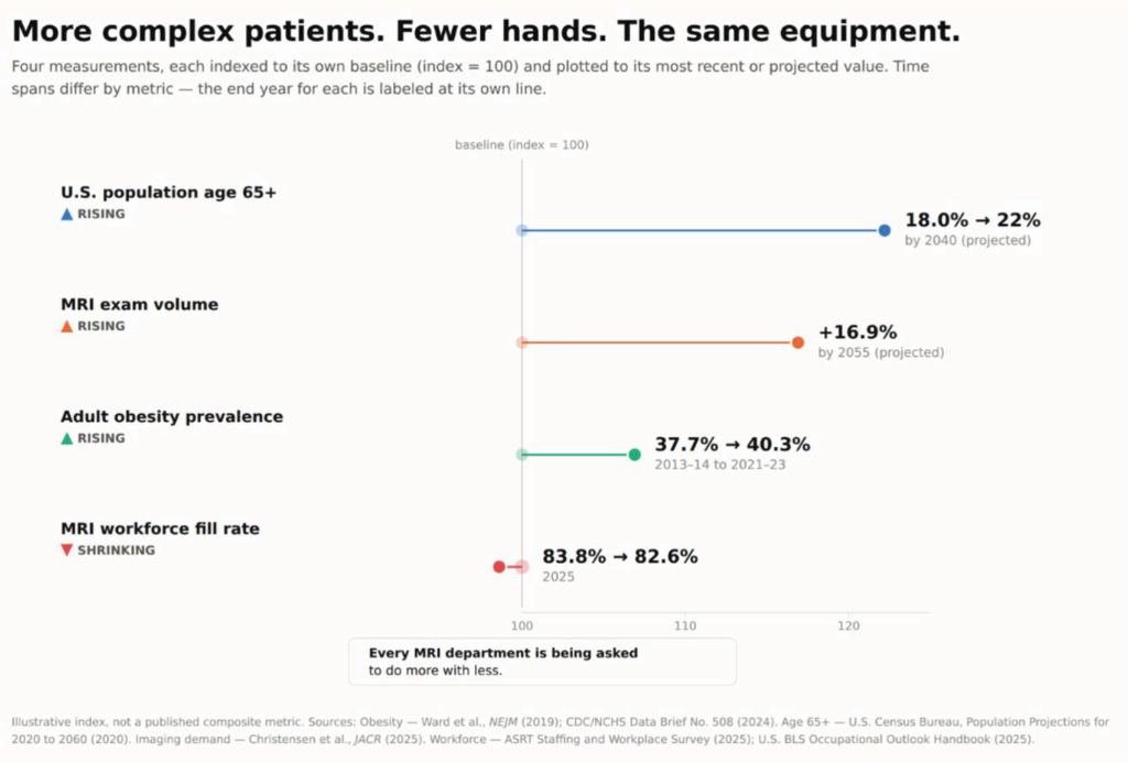
Four pressures are moving at once: an older population, rising MRI demand, high adult obesity prevalence, and increasing MRI workforce vacancy rates. Source details are included in the figure.
The Post-Transfer Gap
Non-standardized positioning is needed when a patient does not fit the standard setup—when body habitus, spinal alignment, or mobility means the technologist must build and maintain a stable position rather than simply place the patient in a coil. An estimated 20–40% of adults over 60 have measurable hyperkyphosis, and national adult obesity prevalence exceeds 40%. These measures do not represent the share of MRI patients who need non-standard positioning; they indicate populations for whom a non-standard setup may be more likely.
In practice, this can mean rolled towels along the trunk to limit lateral movement, pillows stacked and re-stacked as they compress under weight, or a technologist holding a limb or head in place because nothing else will keep it still. These materials were not designed to bear a patient’s weight throughout a full protocol; every adjustment can add motion risk, setup time, and the possibility that the position will not hold through the next sequence.
The distinction from transfer is important. Safe patient handling has standardized many ways to get a patient to the table. The separate challenge begins after transfer: providing reliable lifting and load-bearing support to establish and maintain position in the bore.
The impact often goes unmeasured. In a 2020 study of medical imaging and radiation therapy professionals, 62.6% of the 401 respondents reported experiencing an occupational injury. Among respondents who reported an injury, 81.7% reported muscular injuries. These figures are imaging-wide and do not isolate MRI positioning. Facilities may track the injury itself but often lack a standard way to record how long a positioning challenge took, how much assistance it required, or whether support held throughout the exam.
Turning What You See into a Conversation
Most of it never gets said out loud. It is absorbed by the technologist who finds another way, the exam that runs long, and the shift that ends late.
Technologists can help make the pattern visible—not by escalating every difficult case, but by connecting what happens at the table to the measures leadership already tracks.
- Bring patterns, not isolated incidents. “This is the third time this month I have needed a second person for a similar positioning challenge” can start a useful conversation with a lead technologist or manager.
- Translate the workaround into operational terms. Discuss added setup time, repeat sequences, callbacks, overtime, and staff strain—not only the workaround itself.
- Use existing safety channels. Patient positioning in Zone IV may fall within safe patient handling and mobility (SPHM), safety, or risk-management discussions, even when it has been easy to overlook.
This does not require making a case for a specific product. It starts with naming what is already happening consistently enough for leadership to see the pattern.
When Positioning Becomes the Barrier
Patient story and images shared with permission.
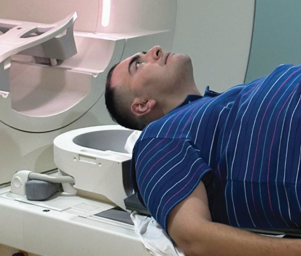
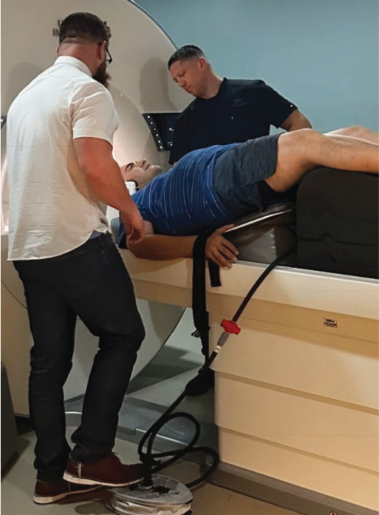
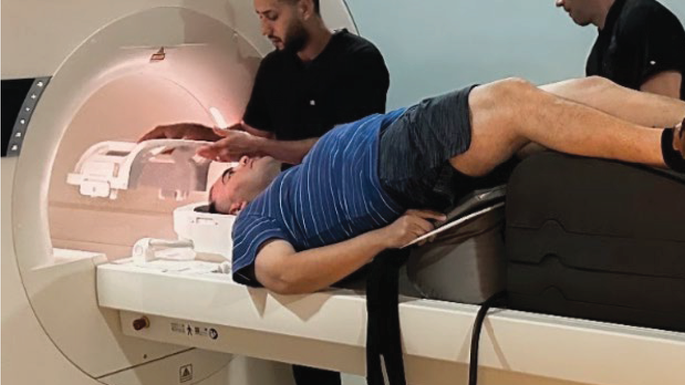
Bobby had been turned away three times before this attempt. Getting him to the table was not the problem. Without additional support, his head and neck remained too far forward for the position required for his exam—a challenge that can occur when kyphosis affects alignment with a standard coil setup.
Instead of relying on manual holding or improvised support that can shift or compress, a load-bearing platform provided a stable base beneath Bobby, helping maintain support and limit side-to-side movement throughout the scan.
A Purpose-Built Approach to Positioning
The goal is not to redefine every positioning challenge as a KyphoLift problem. It is to recognize a category of handling exposure that sits between established SPHM processes and routine imaging workflow.
KyphoLift was developed for this part of the MRI workflow: providing a repeatable, load-bearing patient-support option in the MR environment when standard positioning is not enough. Facilities should determine whether it is appropriate for their patients, equipment, and clinical workflow.
Facilities should independently review the manufacturer’s labeling, Instructions for Use, field-strength requirements, conditions of use, and their own safety policies before implementation.
Learn more about KyphoLift® → https://mriequip.com
The Next Step in MRI Consistency
Much of MRI has been optimized for today’s growing demands. Patient positioning in Zone IV deserves the same attention. Not every positioning challenge prevents an MRI from being completed, and many will never be formally documented. But the accumulated effects still matter. For technologists, this is not a new problem—it is one they have been solving quietly, at the table, every day. The transfer gets the patient to the table. Positioning determines what happens from there.

2026 Innovation Award Winner
The Innovation Showcase presents Annual Meeting attendees with new or recently updated products, services, and solutions advancing the future of medical imaging.
AHRA was thrilled to present the 2026 award to KyphoLift®!
Supporting research:
KyphoLift Peer-Reviewed Research –
Zone IV Positioning Risk
(Click or Scan the QR Code for PDF)
KyphoLift 2026 WhitePaper – Unrecorded Positioning
Risk Between Transfer and Completed Care
(Click or Scan the QR Code for PDF)
Leave a Reply